The larvae
Friends, I am so immensely proud of my team for our work with Porites lobata coral larvae. I just have to show off some of my favorite photos.
 |
| Porites lobata eggs! Matthew and I were both very excited about the "fat rainbow." It indicates the eggs are full of lipids and were well-provisioned by their mom. |
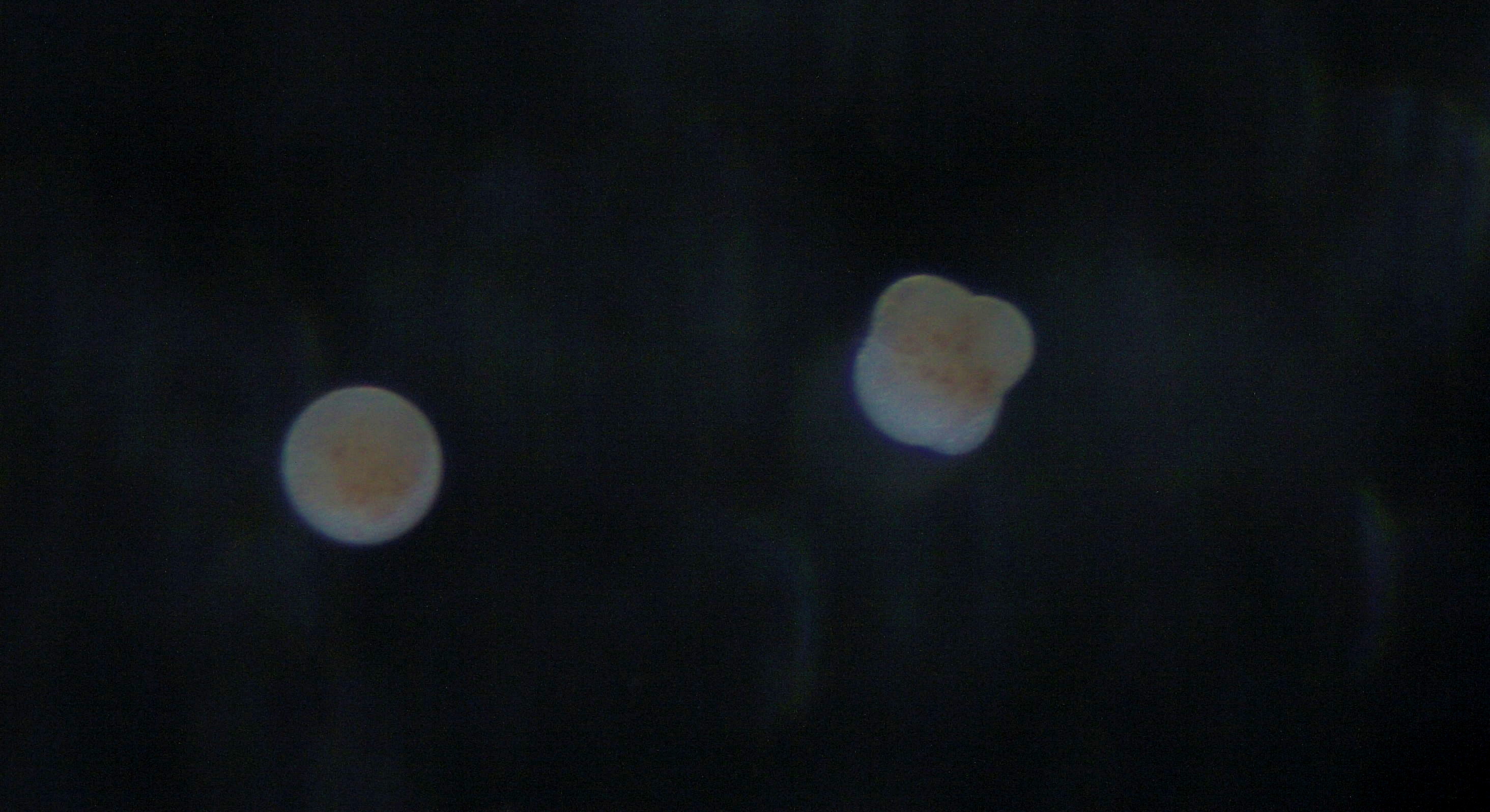 |
| On the left is an egg that has not begun dividing yet. On the right is one that's undergone two divisions and is in its 4-cell stage. |
 |
| Dividing embryos! These are all from the same batch but might have been fertilized at slightly different times, causing them to be at different stages of cell division. I was really confused by the 8- and 16-cell stages in some embryos looking like clusters of grapes rather than organized spheres, but the larvae all seemed to develop normally. |
 |
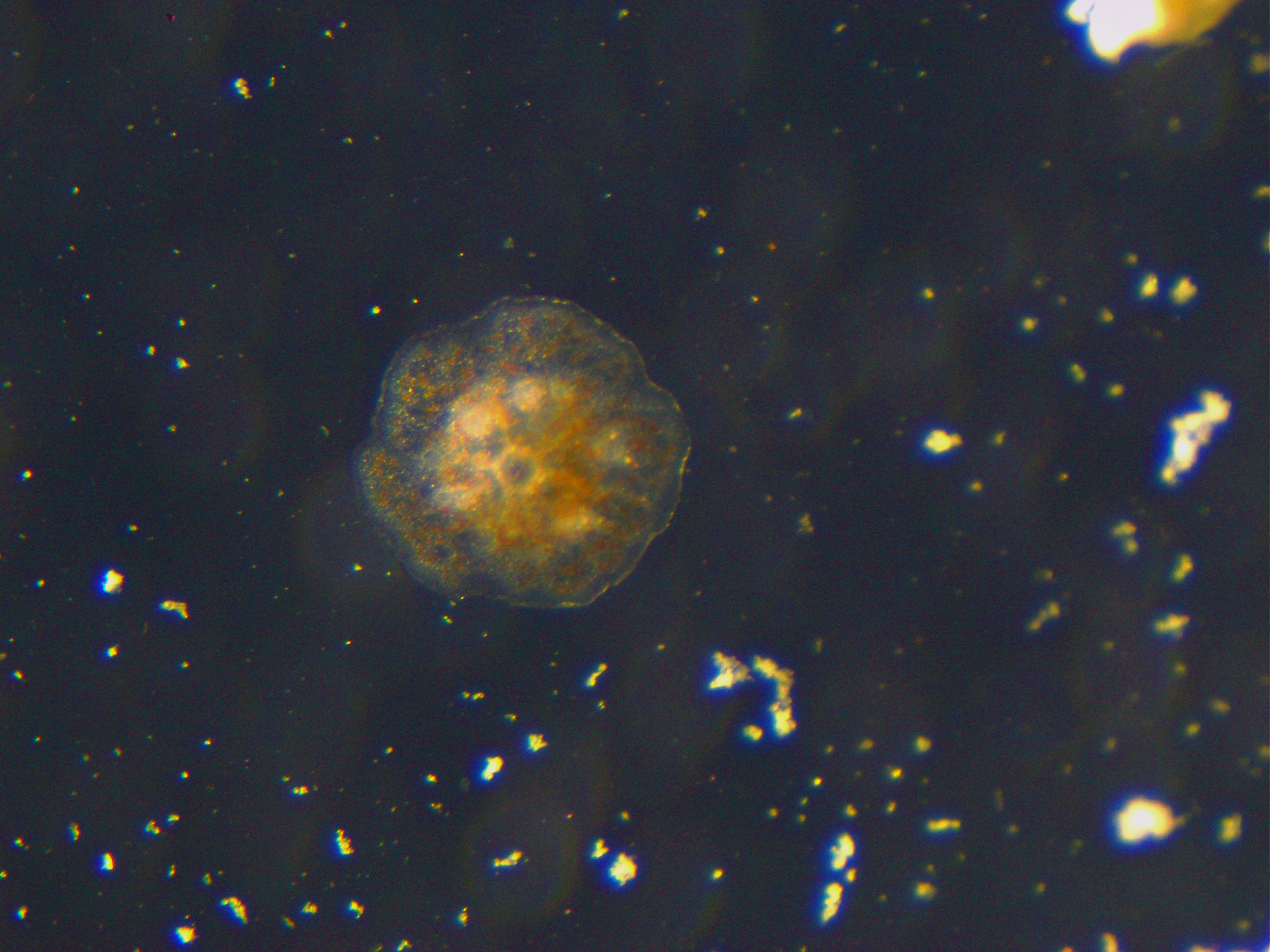
| Swimming larvae! They're slightly elongated and swim with cilia. In this photo, you can even see the symbionts. Those dark brown dots on each larva are zooxanthellae that they inherited from their mom. |
 |
| We discovered that the larvae fluoresce! This picture was taken using a blue light and a yellow filter. |
 |
| This is one of the larvae mid-metamorphosis on a limestone tile. |
 |
| Look at that metamorphosed coral juvenile! This one has just settled on a glass microscope slide. |
 |
| Aah, such a cute baby coral settler! You can see the septa, the mouth, and maybe even the beginning of calcification! |
Comments
Post a Comment